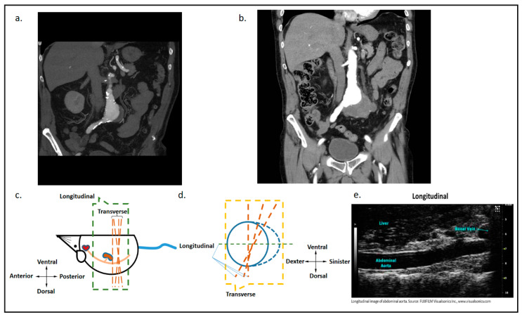
Figure 2.
Coronal images of computed tomographic angiograms of a patient with an AAA and visualisation of an AAA in a mouse model. (a) The image shows a coronal slice of a computed tomographic angiogram of an intact AAA measuring a maximum diameter of approximately 47 mm. (b) One year later the, computed tomographic angiogram showed a retroperitoneal leak from the AAA. The patient underwent successful emergency endovascular aneurysm repair. (c) An illustration of the longitudinal and transverse planes when scanning a mouse aorta using ultrasound in the longitudinal axis. (d) An illustration of longitudinal and transverse planes when scanning a mouse aorta using ultrasound in the transverse axis. (e) A longitudinal plane image of a mouse aorta using micro high-resolution ultrasound. Image source: application brief: abdominal aortic aneurysm, Fujifilm Visualsonics Inc. Permission was obtained to reproduce the image in this manuscript.

