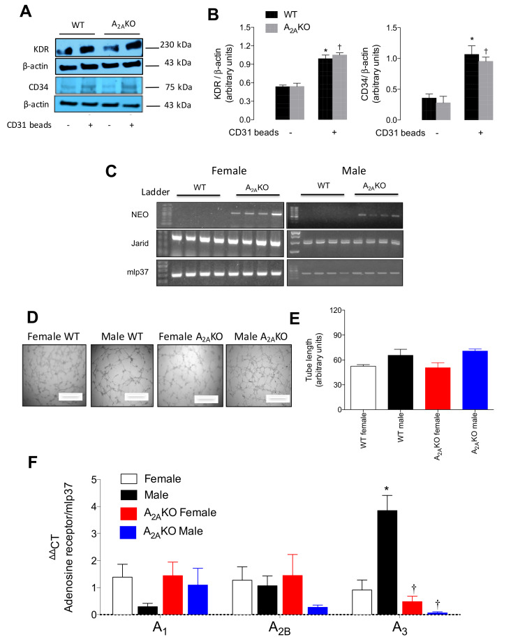
Figure 2.
Expression of adenosine receptors in female and male pulmonary endothelial cells (mPEC) isolated from WT and A2AKO mice. Pulmonary endothelial cells were isolated using collagenase-mediated tissue digestion and immune-selection with CD31-coated Dynabeads. (A) Representative blots of endothelial cell markers. VEGF receptor 2 (KDR, ~230 kDa), CD34 (~80 kDa) and β-actin (~43 kDa) were identified in cell extraction of immunoselected (CD31+, Waltham, MA, USA) cells derived from wild type (WT) and A2A-deficient mice (A2AKO). Cells that were immunoselected are identified with a plus sign (+). (B) Semiquantitative densitometry of KDR/β-actin and CD34/β-actin ratio. (C) Confirmation of gender and genetic background of mPEC isolated from females (single band in the PCR for Jarid gene) or male mice (double band). A2AKO cells were identified by positive amplification of neomycin cassette (NEO). mlp37 gene was used as housekeeping. DNA Ladder 100 bp. (D) In vitro angiogenesis assay at 4 h of incubation with bovine serum (1%). (E) Quantification of tube length of angiogenesis in vitro. (F) QPCR analysis of mRNA levels of A1, A2B, and A3 adenosine receptors in male and female WT and A2AKO mice. See Table S1 for details about primers and PCR amplicons. In (B) * p < 0.05 versus CD31- cells in WT mice. † p < 0.05 versus CD31- cells in A2AKO. In (F). * p < 0.05 versus respective value in female WT mice. † p < 0.05 versus respective value in male WT mice. Values were expressed as mean ± SEM. n = 3–5 per group. All experiments were performed in duplicate.

