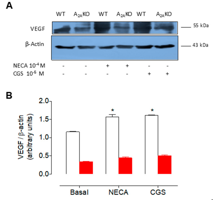
Figure 6.
Expression of VEGF in female mPEC derived from wild type and A2AKO mice. (A) Representative Western blot of VEGF (~55 kDa) and β-actin (~43 kDa) in mPEC from female WT (white bars) and A2AKO mice (red bars). Cells were incubated (12 h) with (+) or without (-) NECA (10−4 M) or CGS-21680 (10−5 M). (B) Semiquantitative densitometry of VEGF/β-actin ratio as in (A). * p < 0.05 respect to basal. Values are mean ± SEM, n = 4 per group in duplicates.

