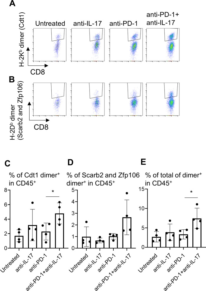
Figure 7.
Therapy combining anti-interleukin-17 (anti-IL-17) with anti-PD-1 increases neoantigen-specific CD8+ T cell infiltration into YTN16 tumors. YTN16 tumor cells were inoculated into four mice as in figure 5. On day 20, tumors were harvested, and tumor-infiltrating cells (TICs) were stained with MHC class I dimer complexed with neoantigen peptides. (A) Dot plots show H-2Kb-restricted mCdt1-specific CD8+ T cells and (B) H-2Db-restricted mScarb2-specific and mZfp106-specific CD8+ T cells. (C) Bar graphs show the percentages of H-2Kb-dimer+ CD8+ T cells in CD45+ TICs. (D) The percentage of H-2Db-dimer+ CD8+ T cells in CD45+ TICs. (E) The sum of these dimer+ CD8+ T cells in CD45+ cells. *p<0.05, Student’s t-test between anti-PD-1 and anti-PD-1+anti-IL-17. MHC, major histocompatibility complex; PD-1, Programmed cell death protein 1.

