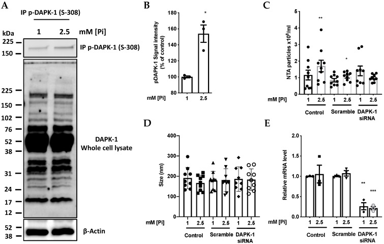
Figure 3.
Pi induces an increase in phosphorylation of DAPK-1. (A) Endogenous human DAPK-1 was immuno-precipitated from cell lysates (200 µg total cellular protein) from cells treated with 1 or 2.5 mM Pi for 1.5 h. Immuno-precipitated DAPK-1 was subjected to immuno-blotting with phosphospecific antibody against p-(S308)-DAPK-1. (B) Densitometry analysis of (A), n = 3, * p = 0.037. (C) Partial silencing of DAPK-1 blunts the effect of elevated Pi concentration on MV release detected by NanoSight Nanoparticle Tracking Analysis (NTA). Each pair of data points refer to a separate multiwall plate, n = 4, * p = 0.015, ** p = 0.0035, (D) Size of particles released from cultures in response to Pi-loading for 1.5 h as in (C) measured by NTA. (E) Relative mRNA level of DAPK-1 confirming siRNA silencing of DAPK-1as indicated in (B–D), *** p = 0.0002.

