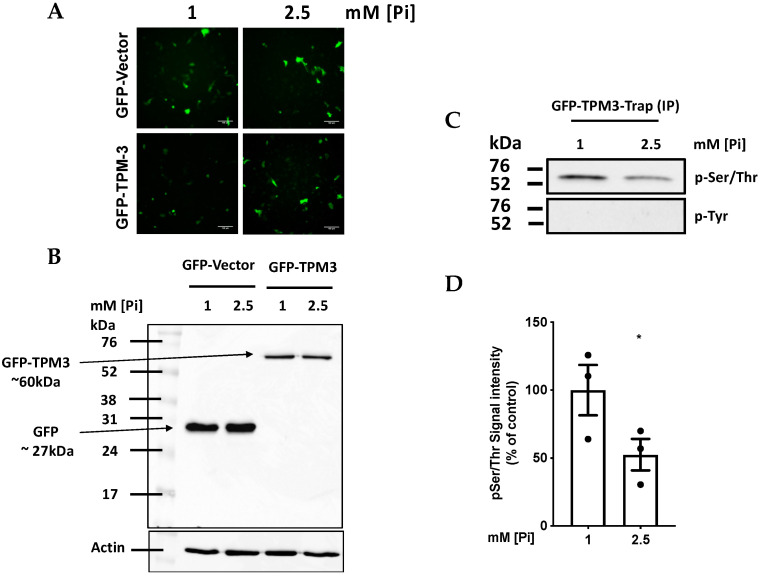
Figure 4.
High Pi induces hypophosphorylation of TPM-3 on a Ser/Thre residue. ECs were transfected with empty GFP-vector or GFP-tagged TPM-3. Thereafter cells were treated with 1 vs. 2.5 mM [Pi] for 1.5 h. (A) Fluorescence microscopy confirming that the cells were transfected successfully. (B) Immuno-blotting using anti-GFP antibody confirming the expression of free GFP (lanes 1 and 2) and TPM-3-GFP (lanes 3 and 4). (C) Immuno-blotting using pan-specific antibodies against pSer/Thr (upper panel) and pTyr (lower panel) demonstrating that GFP-trapped TPM-3 from 200 µg total cellular protein is hypophosphorylated on Ser/Thr but not Tyr in cells treated with 2.5 mM Pi (D) densitometry analysis of (C), n = 3, * p = 0.033.

