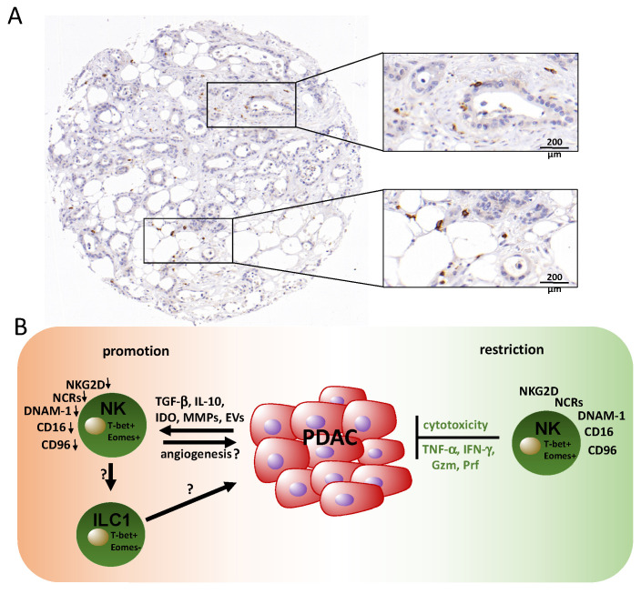
Figure 2.
Natural killer cells and their roles in PDAC. (A) Immunohistochemical staining of CD56 reveals disseminated natural killer (NK)/NKT (Natural killer T-cells) cells (brown). The depicted case shows a rather high density of NK cells considering the distinct heterogeneity of NK cell infiltration, which is generally low in PDAC tissue. (B) PDAC NK cells isolated from the periphery are characterized by a reduced expression of cytotoxicity receptors and exhibit impaired anti-tumor activity, which is induced by mediators of the tumor microenvironment. Restoration of NK cell functions is possible, e.g., via ex vivo activation with IL-2 and gamma-irradiated feeder cell lines. The phenotype of tumor-infiltrating NK cells or other innate lymphocytes such as ILC1 cells and their activity is poorly understood. Gzm: granzyme; Prf: perforin.

