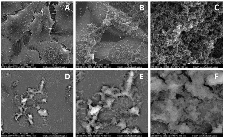
Figure 1.
Scanning electron microscopy (SEM) micrographs of MG-63 cells cultured during 24 h: control (A) and in the presence of 500 ppm doxorubicin-conjugated iron oxide nanoparticles (NP-DOX) (B–F). Information was acquired from secondary electrons (A–E), respectively back scattered electrons (D–F). Images are acquired at different magnifications: 1000× (D), 2000× (E), 4000× (A,B), 5000× (F) and 100,000× (C).

