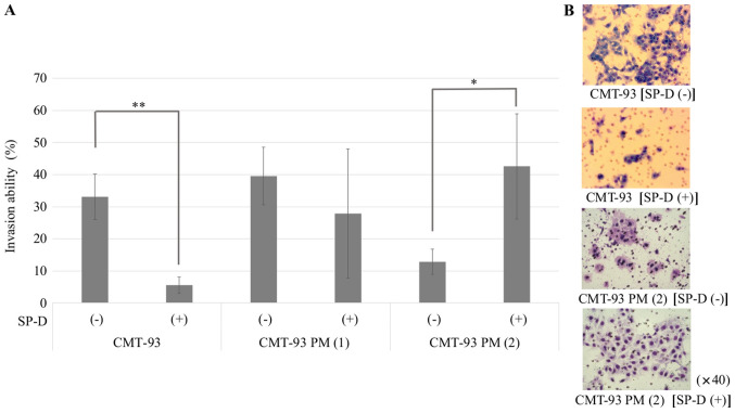
Figure 9.
Cell invasion assay of CMT-93 cells and CMT-93 PM using SP-D. (A) Both CMT-93 PM cell lines showed resistance to treatment with 10 µg/ml of the SP-D protein, whereas SP-D suppressed the invasion ability of CMT-93 cells. CMT-93 PM (2) enhanced the invasion capacity due to the SP-D treatment (P=0.016). The data shown are mean ± SD. *P<0.05, **P<0.01 compared with the control. (B) Representative images of the stained CMT-93 cells (purple) that passed through the membrane treated without SP-D and with SP-D, and images of the stained CMT-93 PM (2) cells (purple) that passed through the membrane treated without SP-D and with SP-D. SP-D, surfactant protein D; CMT-93 PM, CMT-93 pulmonary metastases.

