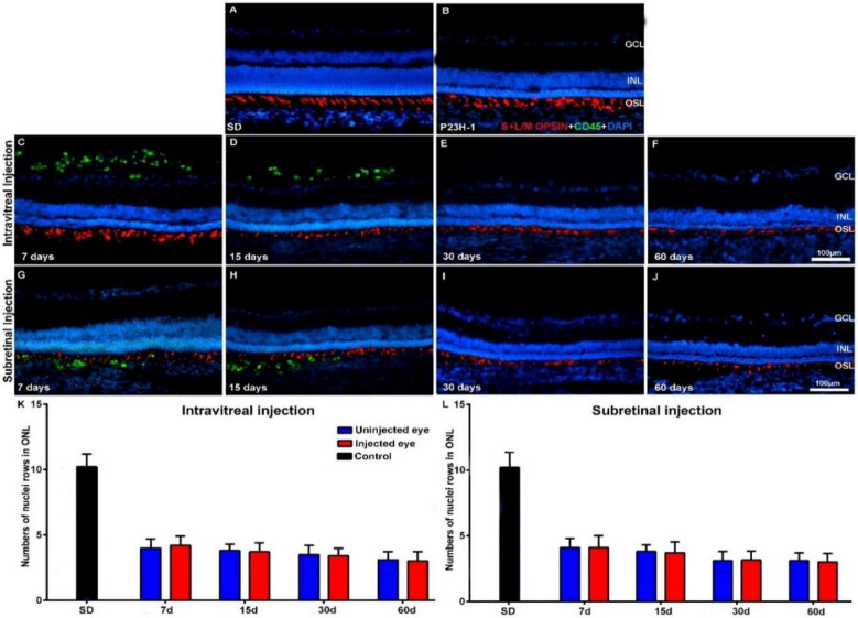
Figure 2.
Cone morphology and ONL thickness in transplanted P23H-1 rats. Microphotographs of representative retinal cross-sections taken from control Sprague-Dawley (SD) rats (A), and the right untreated eyes (B) and left treated eyes (C–J) of P23H-1 rats that received IVI (C–F) or SRI (G–J) of hBM-MSCs. Immunostained cones (red; rabbit anti-red/green opsin and goat anti-blue opsin antibodies) and transplanted cells (green; mouse anti-human CD45 antibody) and also DAPI counterstaining (blue) of the retinas can be observed at different time periods after the injection. Graphs show the mean numbers ± SD of nuclei rows in the ONL of control SD rats (black bars; include data from both right and left eyes) and in the right uninjected eyes (blue bars) and left eyes (red bars) of P23H-1 rats that received IVI (K) or SRI (L) of hBM-MSCs. No significant differences were observed between right and left eyes at any period. n = 6 eyes for control group and treated eyes group at all time points studied; n = 12 eyes for untreated eyes group at all time points studied. Scale bar: 100 μm. GCL = ganglion cell layer; INL = inner nuclear layer; OSL = photoreceptors outer segment layer.

