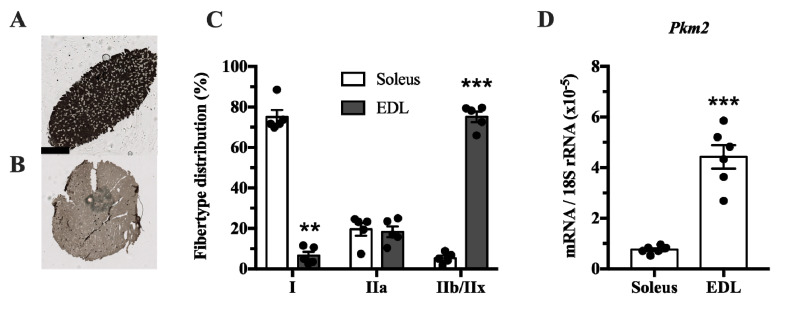
Figure 1.
Pkm2 expression is highest in extensor digitorum longus (EDL) rat muscle that predominantly consists of type 2 fibers. (A,B) ATPase staining of (A) soleus and (B) EDL rat muscle to determine fiber types (n = 4). (C) Fiber type distribution of rat soleus and EDL muscle. (D) Pkm2 mRNA expression in rat soleus and EDL (n = 6). Black circles indicate individual data points. * Significantly different from control, unpaired t-test and Mann–Whitney U test (p < 0.05); ** (p < 0.01); *** (p < 0.001). Scale bar in A indicates 1000 µm.

