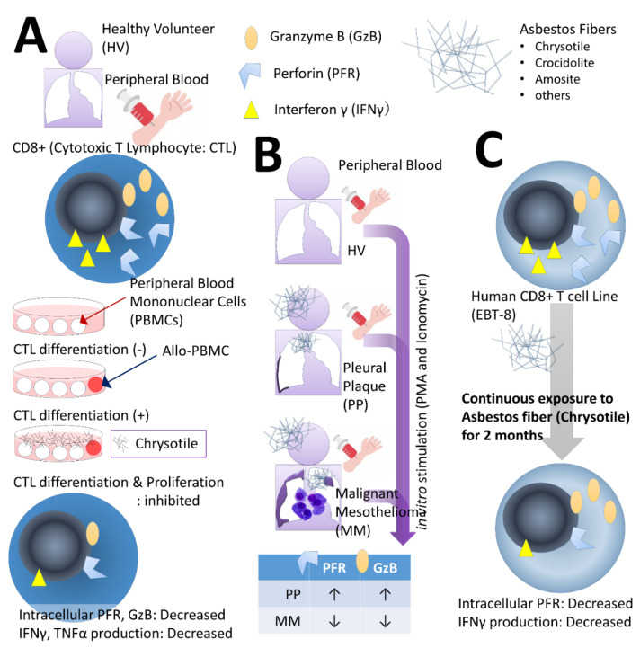
Figure 1.
Observation of the effect of asbestos fibers on CD8+ cytotoxic T lymphocytes (CTLs). In the peripheral blood mononuclear cells (PBMCs) collected from (A) healthy volunteers (HVs), clonal expansion of CTLs was observed in the mixed lymphocyte reaction (MLR). When co-cultured with allogenic PBMCs, CD8+ cells differentiated into CTLs and proliferated, but when chrysotile and asbestos fibers were added, both differentiation and proliferation were suppressed. Furthermore, expression of granzyme B (GzB) and perforin (PFR), which execute the cell killing mechanism, and the production of interferon (IFN)-γ and tumor necrosis factor (TNF)-α, which are closely related cytokines, were also reduced. (B) With HV, CD8+ cells were isolated from the peripheral blood of patients with pleural plaque (PP) and malignant mesothelioma (MM), stimulated with phorbol 12-myristate 13-acetate (PMA) and ionomycin overnight, and expression levels of PFR and GzB in the chamber were observed. In the PP group, both were higher compared with HV and decreased in the MM group. (C) After the human CD8+ cell line ETV-8 was continuously exposed to chrysotile and asbestos fibers for 2 months, intracellular PFR decreased and IFN-γ production decreased.

