Figure 3.
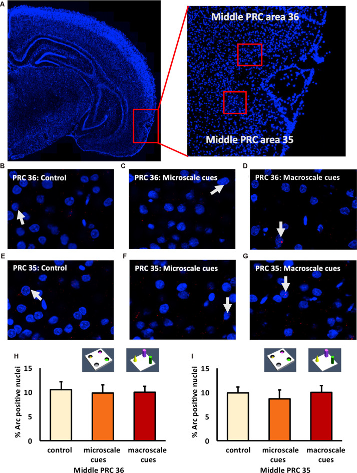
IEG expression in the middle part of perirhinal cortex area 35 and area 36 is unaffected by exposure to novel item-place configurations. (A) The left panel shows a DAPI-stained coronal section (ca. −4.56 mm posterior to Bregma) of the rat brain and the outline of the middle PRC (red square). The right panel shows an enlargement of the outlined area to show the middle PRC in which outlines of area 36 and 35 (red squares) are indicated, where z-stacks were taken for analysis. (B–G) Representative images of nuclear Arc mRNA positive nuclei (red dots, indicated by white arrows) in the middle PRC, area 36 (B–D) and PRC area 35 (E–G) following exploration of microscale cues (C,F), or macroscale cues (D,G) compared to controls (B,E). Blue: nuclear counterstaining with DAPI. Images were obtained using a 63× objective. (H,I) Bar charts describe the relative percentage of nuclei expressing Arc mRNA in the middle PRC after novel item-place exploration, compared to responses detected in control animals (mean ± SEM). No significant changes in nuclear Arc mRNA expression occured in PRC area 36 (H) or area 35 (I) in both experimental conditions, compared to controls (ANOVA; middle PRC area 36 F(2,19) = 0.0630, p = 0.939180; middle PRC area 35 F(2,19)= 0.2444, p = 0.785604).

