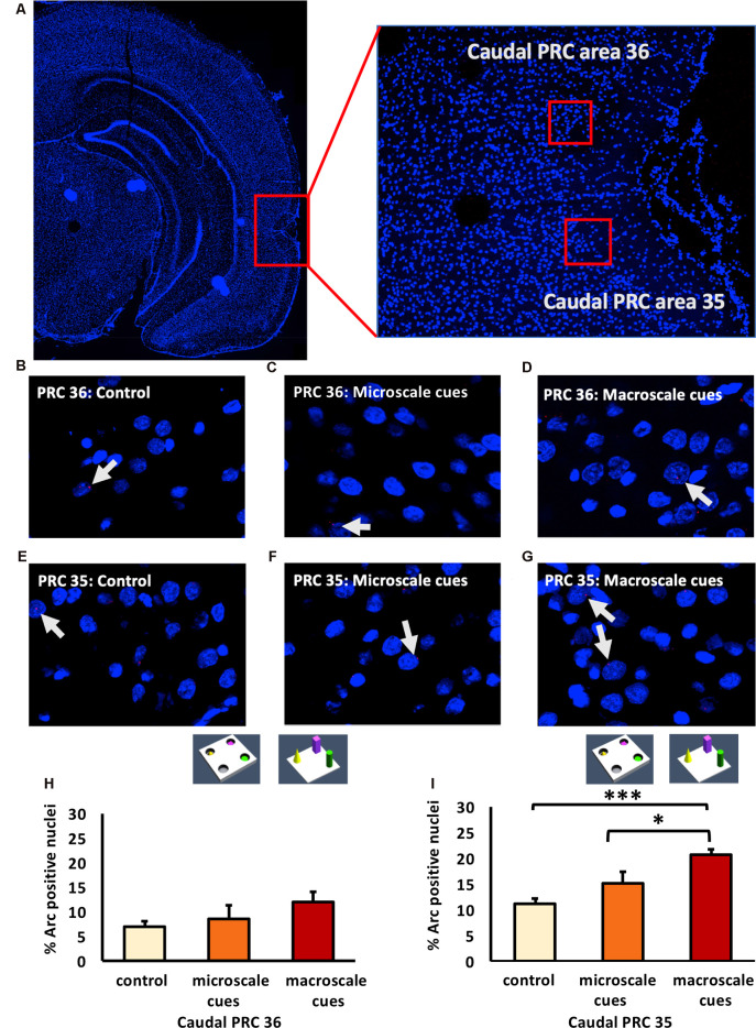
Figure 4.
Exposure to a novel macroscale item-place configuration induces an increase in IEG expression in the caudal part of perirhinal cortex area 35, but not in area 36. (A) The left panel shows a DAPI-stained coronal section (ca. −5.52 mm posterior to Bregma) of the rat brain with the caudal PRC highlighted by a red rectangle. The right panel shows a magnification of the caudal PRC area. Z-stacks were created in the caudal PRC area 36 and 35, indicated by red squares. (B–G) Photomicrographs, taken using a 63× objective, show nuclear Arc mRNA expression (red points, indicated by white arrows) in the caudal PRC (B–D) area 36 and (E–G) area 35 following (C,F) microscale and (D,G) macroscale item-place exploration, or IEG expression in corresponding brain sections from control animals (control; B,E). Cell nuclei (blue) are stained with DAPI. (H–I) Bar charts represent the mean percentage (mean ± SEM) of Arc mRNA positive nuclei in caudal PRC area 36 (H) or PRC area 35 (I) under control or test conditions (ANOVA; caudal PRC area 35: F(2,21) = 7.9498, p = 0.002689; area 36: F(2,20) = 1.18071, p = 0.327571). Exploration of microscale item-place cues does not lead to significant changes in Arc mRNA expression in caudal PRC areas 35 and 36, relative to controls (microscale vs. control, post hoc Fisher’s LSD test, p > 0.05, each). Caudal PRC area 35, but not area 36, responds to macroscale item-place information (post hoc Fisher’s LSD test; area 36 p > 0.05; area 35: ***p < 0.001 for macroscale vs. control and *p < 0.05 for macroscale vs. microscale).

