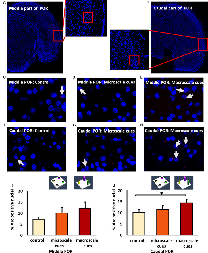
Figure 5.
Differentiated IEG expression in the middle and caudal parts of the postrhinal cortex (POR) following the exploration of novel item-place configurations. (A,B) DAPI-stained coronal sections of the rat brain show the regions of interest examined in the (A) middle and (B) caudal parts of the POR, as indicated by red squares and the respective magnified images (middle). The red squares in enlarged images indicate areas where z-stacks were obtained for the middle POR (ca. −6.96 mm posterior to Bregma) and for the caudal POR (ca. −7.8 mm posterior to Bregma). (C–H) Photomicrographs represent nuclear Arc mRNA expression (red dots, indicated by white arrows) in the middle POR (C–E) and caudal POR (F–H) of control animals (C,F) or animals that participated in microscale (D,G) or macroscale item-place exploration (E,H). Nuclei (blue) were stained with DAPI. Images were taken using a 63× objective. (I,J) Bar charts showing the relative percentage of positive Arc mRNA nuclei in the middle POR (I) and caudal POR (J) of controls and for both experimental conditions (mean ± SEM). No significant changes can be observed in the middle POR (I) following microscale and macroscale item-place exploration compared to the control group (ANOVA F(2,19) = 1.19376, p = 0.324814). Interestingly, in caudal POR (J), a significant difference in Arc mRNA expression can be detected in animals that participated in macroscale item-place exploration compared to the control group (post hoc Fisher’s LSD test, *p < 0.05), whereas exposure to microscale cues does not change Arc mRNA expression (p > 0.05).

