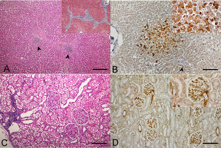
Fig 3.
Domestic cat hepadnavirus infection of the (A, B) liver and (C, D) kidney. Representative (A, C) H&E and (B, D) DHC IHC images from cat no. 3. (A) Hepatic fibrosis with sinusoidal lymphocytic infiltration (arrowheads) plus positive staining to Masson-Trichrome special staining (inset). Bar indicates 45 μm. (B) DCH-immunoreactivity signals were indicated in cytoplasm of hepatocytes (inset) in the area adjacent to hepatic fibrosis (arrowhead). Bar indicates 170 μm. (C). Membranoproliferative glomerulonephritis with focal interstitial nephritis. Bar indicates 170 μm. (D) DCH immunopositivity was observed in the vascular pole, glomerular capillary loops and basement membrane, and mesangial cells (inset). Bars indicate 170 μm.

