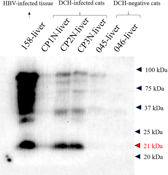
Fig 6. Cross-reactivity of anti-HBcAg with the DCH.
Western blot analysis revealed positive reactivity of 21 kDa protein of DCH-positive liver samples (CP1N-CP3N-liver) that was similar to result of the HBV-positive sample (158-liver). No reactivity was observed in DCH-negative samples (nos. 045- and 046-liver).

