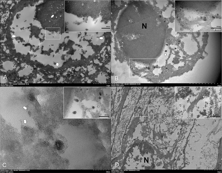
Fig 7. Ultrastructure of DCH and intracellular development of DCH particles in the liver.
Representative TEM images from the liver of cat no. 3. (A) The DCH antigen spheres (inset) were detected in the nucleus of hepatocytes. Arrows indicate the nuclear membrane. (B) Numerous DCH particles were found in the nucleus (N) with a single viral sphere inside the nuclear membrane (asterisks). A complete Dane-like particle (arrow) was observed in the cisternae of the ER and numerous incomplete Dane-like particles and viral particles were attached to the ER membrane (inset). (C) Invagination of the ER membrane with a complete Dane-like particle. Incomplete Dane-like particles stalked with the ER membrane and attached viral particles with the ER membrane were observed (arrows). The Dane-like particles were frequently observed as free-floating and attaching to the membrane (inset). (D) Numerous clusters of DCH core particles (inset) in the cytoplasm of fibrous cells residing in the area of fibrosis. Bars indicate as described in figures.

