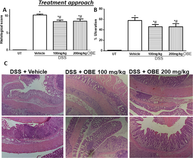
Fig 2. Effect of OBE on colitis severity at the histological level using the treatment approach.
The histological assessment of colitis severity (panel A), and the % of ulceration in the whole colon section (panel B) were determined in mice receiving DSS and either various doses of OBE (hatched bars) or vehicle (solid bars), and in the UT healthy mice (open bars). Histobars represent means ± SEM for the following number of mice in each group: UT (n = 10), DSS/vehicle (PBS) (n = 16), DSS/OBE 100 mg/kg (n = 20), and DSS/OBE 200 mg/kg (n = 12). Asterisks denote significant difference from UT mice with p<0.05 (*), # denotes significant difference from DSS/vehicle-treated mice with p<0.05. Panel C (left side) is an illustration of a colon section taken from mouse treated with DSS/vehicle where there is significant mucosal destruction, submucosal edema formation, increase muscle thickness, and significant immune cells recruitment. The middle and left sides are illustrations of typical colon sections taken from mice treated with 100–200 mg/kg doses of OBE. There was a slight improvement in the mucosal integrity and slight reduction in immune cells recruitment (10x magnification).

