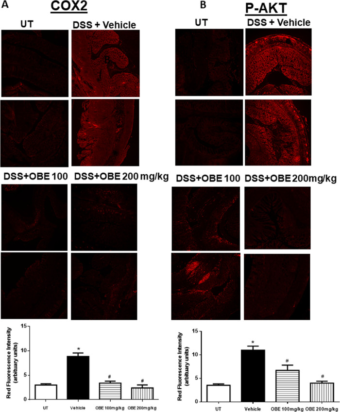
Fig 7. Effect of OBE on colonic COX-2 expression and AKT phosphorylation levels using the preventative approach.
Colon sections taken from mice treated with DSS plus either vehicle (solid bar) or OBE (100–200 mg/kg, hatched bar) or untreated (UT, open bar) mice were immunostained with antisera against COX-2 (A), and phosphorylated AKT (B). Immunofluorescent (Alexa Fluor) signals shown in left side. Histobars (right side) represent quantitative assessment of fluorescence intensity (arbitrary units), and represent means ± SEM for 4 mice in each group. Asterisk denotes significant difference from the UT mice, with p<0.05, and # denotes significant difference from DSS/vehicle-treated mice, with p<0.05.

