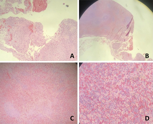
Figure 4. Photomicrograph of the upper pole and the lower pole showing the following aspects: A. HE x10: fibrosis area; B. HE x20: area of fibrosis with foreign body reaction; C. HE x20: necrosis area; D. HE x40: necrosis area with hemosiderophagus. Note that the white pulp and red pulp are preserved.

