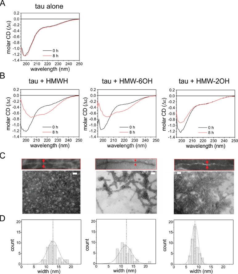
Figure 4.
Secondary structure and morphology of Δtau187 aggregates. (A) CD spectra of Δtau187 (20 μM) at 0 and 8 h. (B) CD spectra of Δtau187 in the presence of 5 μM HMWH, 6-O-desulfated HMWH, or 2-O-desulfated HMWH at 0 and 8 h. The samples were incubated at 37 °C without agitation. All CD spectra are shown as the average of triplicate repeats. (C) TEM images of aggregates formed from Δtau187 (20 μM) after incubating with 5 μM of HMWH or the desulfated derivatives without agitation for 24 h. Scale bar = 200 nm. (D) Distribution of filament widths measured from the TEM images.

