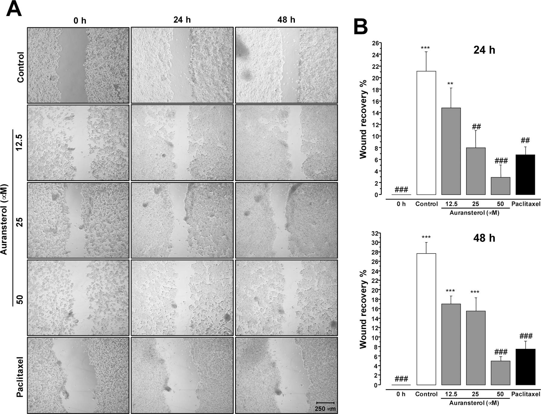
Fig. 5.

Effect of auransterol on HT-29 colon cells migration. (A) Microscopic view (magnifier Zeiss CP-Achromat 5x) of cells were treated 48 h with auransterol (0–50 μM) or paclitaxel (0.01 μM) on wound healing assay. Cell migration was monitored with 5 x magnification. (B) Quantitative analysis of cell migration into the scratch wound at 24 or 48 h post-scratch. Data are expressed as the means ± SEM (n = 3) and analyzed by one-way ANOVA using Dunnett’s multiple-comparison test (*P < 0.05, **P < 0.01, ***P < 0.01 compared to the 0 h; ##P < 0.01, ###P < 0.001 compared to the control) of three independent assays.
