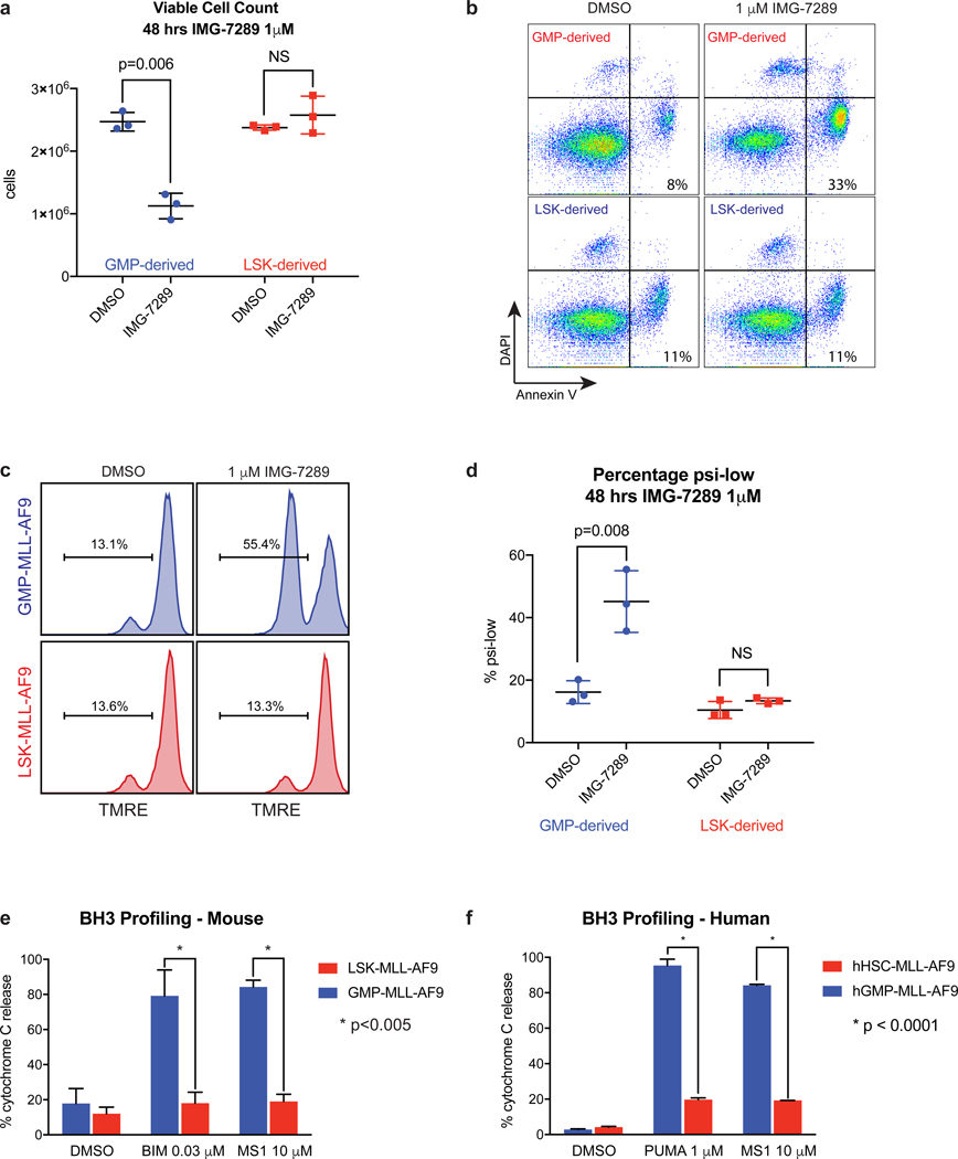
Figure 2. Differential apoptotic thresholds in murine and human MLL-AF9 leukemias arising from distinct cells of origin.
LSK- and GMP-derived MLL-AF9 leukemias were treated with DMSO or 1 μM IMG-7289. (A) Viable DAPI− cells were enumerated after 48 hours of treatment. (B) Annexin V and DAPI staining was assessed by flow cytometry. p<0.03, using Student’s t-test comparing percent Annexin V-positive cells between GMP- and LSK-derived. Data shown are representative of 3 independent experiments. (C) TMRE staining was measured by flow cytometry as a readout of mitochondrial membrane potential after 48 hours of treatment. Statistical analyses summarized in (D). Data shown are representative of 3 independent experiments. (D) Summary scatter plots depicting the percentage of cells within the ψlow gate defined by TMRE flow cytometry in (C). BH3 profiling of (E) murine LSK- and GMP-derived MLL-AF9 leukemia cells or (F) human cord blood HSC- and GMP-derived leukemias exposed to DMSO control or pro-apoptotic peptides.

