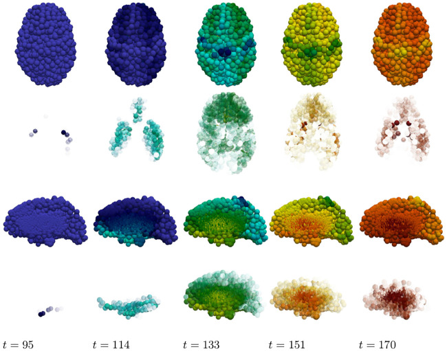
Fig 18. Damage progression in secondary tauopathy.
Horizontal plane view (top row) with opacity exaggerated (second row) progression. sagittal view (third row) with opacity exaggerated (fourth row) progression. The color scale is identical to that of Fig 17.

