Figure 2.

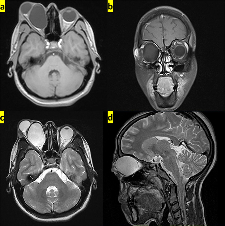
The transverse and coronal planes from MRI show hypointense hydatid cyst on T1-weighted images (a and b), whereas it appears hyperintense in the transverse and sagittal planes from MRI on T2-weighted images (c and d).

The transverse and coronal planes from MRI show hypointense hydatid cyst on T1-weighted images (a and b), whereas it appears hyperintense in the transverse and sagittal planes from MRI on T2-weighted images (c and d).