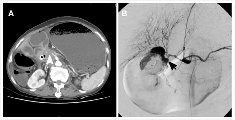
Figure 2.
A 72-year-old patient with pancreatic cancer and duodenal obstruction. (A) Computed tomographic imaging before the procedure reveals a tumor encasing the duodenal bulb. Outline of the celiac and gastroduodenal arteries is irregular due to tumor invasion (white arrowhead). (B) Massive hematemesis is observed 3 days after stenting. Angiography reveals contrast medium extravasation into the duodenum near the stent (black arrow).

