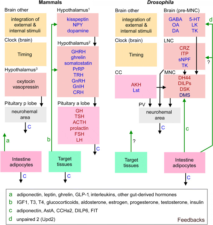
Fig. 13.
Comparison between mammalian neurosecretory systems and those of Drosophila. This highly schematic figure shows the key features of the brain-hypothalamus-pituitary axis in comparison with presumably analog regions in Drosophila. Same colors indicate presumed similar regions. Hypothalamus is divided into three regions (1–3) and the pituitary into anterior (a) and posterior (p) lobes. Substances indicated in blue are neuromodulators and/or release-regulating factors, those in dark red are hormones and the function of dromysosuppressin (DMS) in MNCs is not clear although the DMS-MNC cells have axons terminating together with those of the other MNCs. Arrows indicate action of the substances on cells in a region, blue C:s indicate release into general circulation and green arrows are feedback from target tissues. The regions “brain other” and “clock” are not interconnected to other regions with arrows to avoid complexity but certainly play important roles in regulating neurosecretory systems (and receive feedback from hormonal systems). We lumped together intestine and adipocytes as one set of target tissues and other targets are grouped in target tissues (these are, e.g., gonads, thyroid, adrenals and liver in mammals and gonads and muscles in Drosophila). Adipocytes in Drosophila include other fat body functions (e.g., liver-like and immune functions). Substances used as feedback are indicated by green (a–d) and are listed in the box below (GLP-1, glucagon-like peptide 1; IGF1, insulin-like growth factor 1; T3, triiodothyronine; T4, thyroxine; FIT, female-specific independent of transformer). Acronyms of other factors and hormones are as in Table 1. Other abbreviations: CC, corpora cardiaca; MNC, median neurosecretory cells; LNC, lateral neurosecretory cells; PV, proventriculus region (including aorta, crop duct, and CC)

