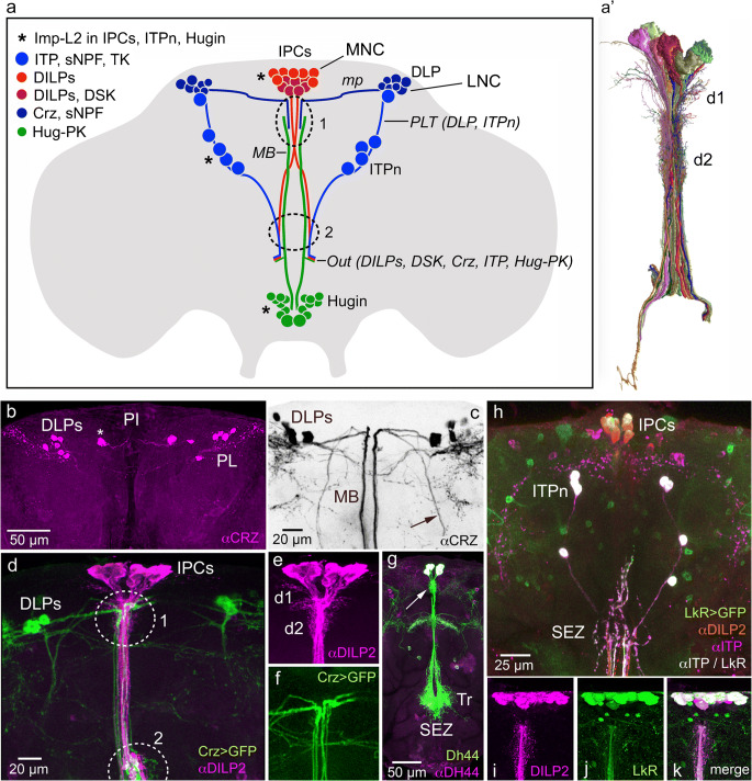
Fig. 3.
Distribution of a selection of peptide hormones in neurosecretory cells in the adult Drosophila brain. (a) Schematic of some of the lateral (LNC) and median (MNC) neurosecretory cell systems in the adult fly brain. Two sets of LNCs are shown: the DLPs that produce corazonin (CRZ) and short neuropeptide F (sNPF) (Kapan et al. 2012) and the ITPn that produce ion transport peptide (ITP), sNPF and tachykinin (TK) (Kahsai et al. 2010). One set of MNCs is depicted, the insulin-producing cells (IPCs) that produce insulin-like peptides (DILP1, 2, 3 and 5) of which a subpopulation also produces sulfakinin (DSK) (Söderberg et al. 2012). A set of neuroendocrine cells (Hugin cells) in the subesophageal zone (SEZ) produces the peptide pyrokinin (Hug-PK); a subset of these constitutes neurosecretory cells (Melcher and Pankratz 2005). Axons from these four cell groups run via the median bundle (MB) or posterior lateral tract (PLT), exit the brain (at Out) via the NCC (nervii corpora cardiaca; not shown) and they reach the retrocerebral complex where axon terminations are in contact with the circulation. The DLPs utilize sNPF as a factor that regulates release of DILPs and AKH (Kapan et al. 2012; Oh et al. 2019) and the ITPn possibly use TK and sNPF as releasing factors and Hugin cells may employ Hug-PK to regulate release of other peptides (Schlegel et al. 2016). Interactions between the cell systems shown probably occur at the sites indicated by 1 and 2. It was shown that three of these cell types (marked with asterisks) express the protein ImpL2 and that the IPCs signal to the ITPn and Hugin cells with DILP2 via the insulin receptor in an Imp-L2-dependent fashion (Bader et al. 2013). mp, medially projecting axons of LNCs. (a’) A set of MNCs (including IPCs) reconstructed from serial electron microscopic sections of Drosophila hemibrain. d1 and d2 two sets of presumed dendrites. This figure was compiled from data in neuPRINT (https://neuprint.janelia.org) (Clements et al. 2020; Xu et al. 2020; Zheng et al. 2018). (b) There are seven pairs of CRZ expressing DLPs in pars lateralis (PL). Note that in this specimen one larger pair of cells (asterisk) is slightly dislocated medially towards pars intercerebralis (PI). (c) The DLPs (inverted image; CRZ immunolabeling, αCRZ) have axons running through the median bundle (MB) and a lateral tract (arrow). (d) The DLPs (green) impinge on the IPCs (magenta) in encircled areas 1 and 2. (e, f) Details of IPCs and branches from DLPs. D1 and d2, two sets of IPC dendrites. (g) Six median neurosecretory cells producing diuretic hormone 44 (DH44) have dendrites at the arrow and axon branches in the tritocerebrum (Tr) and subesophageal zone (SEZ) rendered by Dh44-Gal4-driven GFP and antiserum to DH44. (h) Triple labeling with antisera to ITP (magenta/white), DILP2 (red) and LkR-Gal4-driven GFP (leucokinin receptor; green) reveals ITP neurons (ITPn) and IPCs. This indicates that the LkR is expressed on both ITPn and IPCs. (i–k) The IPCs express the leucokinin receptor (LkR) seen with LkR-Gal4-driven GFP and anti-DILP2. Panel (a) is updated from Nässel et al. (2013) with data from several of the papers cited above. Panel (b) is from Kubrak et al. (2016), (c–f) are from Kapan et al. (2012), (g) from Zandawala et al. (2018a) and (h–k) from Zandawala et al. (2018b), with permission from the publishers

