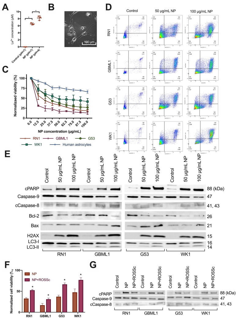
Figure 2.
La2O3 NPs generate La3+ ions and cause cell death in GBM cells via a ROS mechanism. (A) Evidence of increased free La3+ ions in supernatant of 72 h NP solution (1000 µg/mL) at pH = 4 versus pH = 7 versus control (n = 3 per group). Mean differences to control were 6.5 and 8.2 µM respectively. Statistical analysis was performed using student t-test analysis, with significance set at P < 0.05 (*). (B) Light microscopic evidence of vacuolization and cell death in PDCL WK1 after 72 h of exposure to NP (100 µg/mL) at 40 × magnification. (C) Cytotoxicity of La2O3 NPs in PDCLs after 72 h across a concentration range of 0–100 µg/mL with relevance of (D) the apoptotic and autophagic pathways shown in each PDCL and (E) representative western blotting trends indicating intrinsic, extrinsic, and autophagic processes were involved in cell death; cleaved PARP (cPARP), Caspase-9, cleaved Caspase-8 (cCaspase-8), Bcl-2 and Bax; autophagy-related proteins LC3-I and LC3-II; DNA-damage-related ɣ-H2AX. (F) Significantly increased cell viability across all PDCLs 72 h after NP (50 µg/mL) was shown after 1 h pretreatment with radical oxygen species scavenger (ROSSc) with (G) representative western blotting trends showing reduction of cPARP, Caspase-9 and cCaspase-8 changes following pretreatment with ROSSc. Full blots provided in Supplementary. All cell viability data were obtained in technical triplicate over three independent tests and presented as mean ± SD. Statistical analysis was performed using two-way ANOVA analysis, with significance set at P < 0.05 (*).

