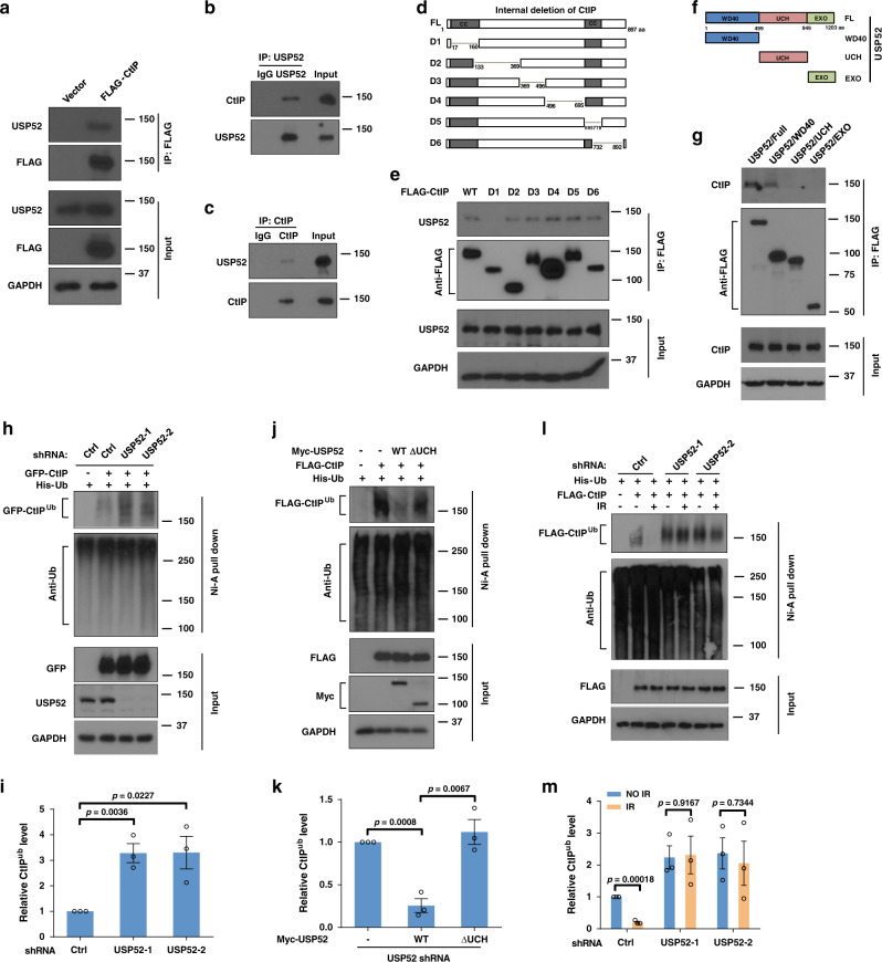
Fig. 1. USP52 interacts with and deubiquitinates CtIP.
a HEK293T cells were transfected with vector or Flag–CtIP for 24 h, cells were then lysed and immunoprecipitated with anti-FLAG agarose beads. The beads were boiled and analyzed with indicated antibodies. b, c HEK293T cell lysates were subject to immunoprecipitation with control IgG, anti-USP52 (b) or CtIP antibodies (c). The immunoprecipitates were then blotted with indicated antibodies. d, e Schematic representation of CtIP constructs used in this study (d). HEK293T cells were transiently transfected with the indicated CtIP constructs for 24 h, then cell lysates were incubated with anti-FLAG agarose beads overnight at 4 °C. The immunoprecipitates were then blotted with indicated antibodies (e). f, g Schematic representation of USP52 constructs used in this study (f). Cellular lysates from HEK293T cells transfected with the indicated constructs of USP52 were immunoprecipitated with anti-FLAG agarose beads, and then western blot was performed with indicated antibodies (g). WD40 WD40 repeat domain, UCH ubiquitin C-terminal hydrolase domain, EXO exonuclease domain. h, i Control or USP52-depleted HEK293T cells were transfected with CtIP and His-Ub for 24 h. Harvested cells were subjected to immunoprecipitation using nickel (His) beads and then the level of ubiquitin conjugates of CtIP was detect by western blotting assay (h). The quantification of bands was analyzed by Image J and data are presented as mean values ± SEM from three independent experiments (i). j, k USP52-depleted HEK293T cells expressing the indicated USP52 constructs were harvested and then immunoprecipitated with nickel (His) beads. Blots were performed to detect the level of ubiquitin conjugates of CtIP (j). The quantification of bands was analyzed by Image J and data are presented as mean values ± SEM from three independent experiments (k). l, m Control or USP52-depleted HEK293T cells were transfected with CtIP and His-Ub for 24 h, cells were then untreated or treated with IR for 2 h. Cell lysates were immunoprecipitated with nickel (His) beads and then blots were performed to detect the level of ubiquitin conjugates of CtIP (l). The quantification of bands was analyzed by Image J and data are presented as mean values ± SEM from three independent experiments (m).

