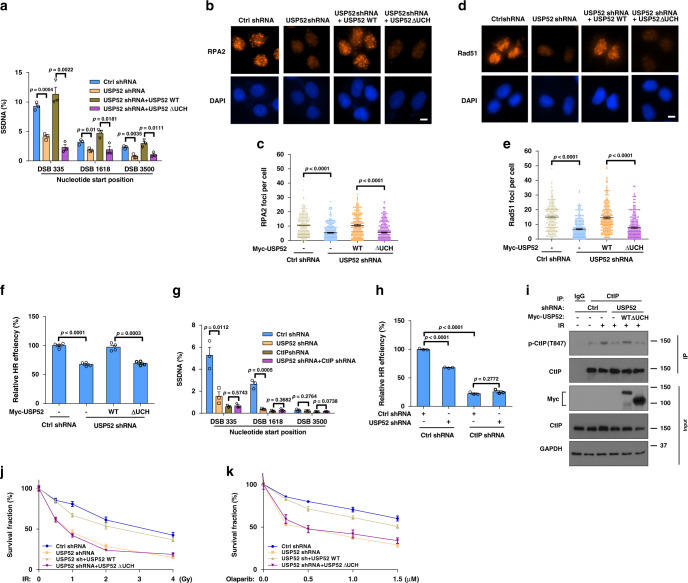
Fig. 3. USP52 regulates DNA end resection and HR through CtIP.
a ER-AsiSI U2OS cells with or without USP52 depletion were transfected with indicated constructs for 24 h, then cells were treated with 1 μM 4-OHT for 4 h before genomic DNA was extracted and digested with BsrGI. DNA end resection efficiency was measured by qPCR assay. Data are presented as mean values ± SEM from three independent experiments. b–e Control or USP52-depleted U2OS cells were transfected with indicated constructs for 24 h before treated with 2 Gy IR for 2 h (b, c) or 5 Gy IR for 5 h (d, e). RPA2 or Rad51 focus formation was then detected by immunofluorescence (b, d) and quantified (c, e). Data are representative of three independent experiments. Each dot represents a single cell, and more than 200 cells were counted in each group for this experiment. Error bars represent SEM from this experiment. Scale bar, 10 μm. f Control or USP52-depleted HEK293T cells reconstituted with the indicated constructs and HR reporter were subjected to HR assay. Error bars represent SEM from three independent experiments. g Control or USP52-depleted ER-AsiSI U2OS cells were transfected with control or CtIP shRNA, and then cells were treated with 4-OHT for DNA end resection assay. Each bar represents SEM from three independent experiments. h Control or USP52-depleted HEK293T cells were transfected with control or CtIP shRNA and HR reporter, and then cells were harvested for HR assay. Error bars represent SEM from three independent experiments. i Control or USP52-depleted HEK293T cells were transfected with WT USP52 or USP52 ΔUCH for 24 h before treated with 5 Gy IR for 1 h. Cells were harvested and subject to immunoprecipitation with control IgG and CtIP antibodies. The immunoprecipitates were then blotted with indicated antibodies. j, k Control and USP52-depleted U2OS cells stably expressing WT USP52 or ∆UCH were in response to the indicated dose of IR (j) or PARPi (k) for 2 weeks. Cell viability was assessed using colony formation assay. Error bars represent SEM from three independent experiments.

