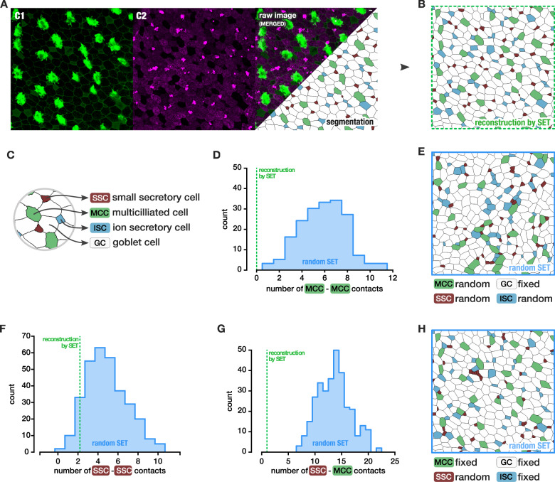
Fig. 4. Cellular organization of the Xenopus tailbud embryo epithelium.
a Image of a st. 33 Xenopus epithelium, actin in membrane and cilia labeled with phalloidin-488 and acetylated α-tubulin-488 (C1, green). Lectin PNA 594-stained mucins to define the four cell types47 (C2, magenta). Scale bar: 10 µm. Segmentation and labeling of four cell types: Multiciliated cells (MCC) are large cells with a high level of actin stain and a lack of lectin PNA. Goblet cells are large cells containing mucins (lectin PNA) and a lack of apical actin. Ionocyte (ISC) are small cells with visible actin and a lack of lectine PNA. Small secretory cells (SSC) are characterized by the absence of actin and dotted lectin PNA highly stained. b Reconstruction of image a by SET, with cell type preserved. c Legend of cell types. d MCC–MCC contacts count observed in the reconstruction by SET (green) compared to the counts obtained in a distribution of SET with random position of the intercalating cells only (blue distribution): p value < 0.006. e An example of SET with random position of the intercalating cells. f SSC–SSC contacts count observed in the reconstruction by SET (green) compared to the counts obtained in a distribution of SET with random position of the SSC only (blue distribution): p value = 0.141. g MCC–SSC contacts count observed in the reconstruction by SET (green) compared to the counts obtained in a distribution of SET with random position of the SSC only (blue distribution): p value < 0.003. h An example of SET where only the small secretory cells positions are reshuffled. e An example of SET with random position of the SSC only. N = 1 image.

