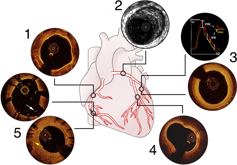
Fig. 1.
Summarizing figure for situations where intracoronary imaging should be considered (Table 2). (1) OCT or IVUS for pre-PCI stent sizing and identification of deployment site, OCT for lesion characterization: OCT image showing severe calcifications. (2) IVUS for ostial left-main lesion assessment and guidance: IVUS image showing left-main plaque. (3) Functional measurement (FFR/iFR) for uncertainty about severity of distal lesions and OCT for composition: iFR/FFR and OCT image showing intimal thickening. (4) OCT for bifurcation lesion assessment and guidance: OCT image showing a bifurcation with high resolution. (5) OCT for post-PCI stent assessment and optimization and assessment of stent failure: OCT images demonstrating malapposition (white arrow shows an intraluminal stent strut) and in-stent restenosis (yellow arrow shows a stent strut covered by neointima hyperplasia)

