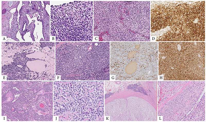
Figure 2. Morphologic spectrum of tumors with EWSR1-CREM fusion including epithelioid, round and spindle cell components.
A, B (case 6, 25/M, intra-abdominal lesion). Cystic metastasis to the liver (A), showing at high power a mixture of primitive round cells with areas of epithelioid cells arranged in solid sheets (B). C, D. (case 11, 14/F, thigh) Solid and cystic soft tissue mass showing epithelioid cells with clear cytoplasm arranged in sheets (C); tumor was diffusely positive for CD99, being misinterpreted as an Ewing sarcoma (D). E-H. (case 12, 29/M) Cystic and solid renal tumor (E) composed of predominantly round cell with focal areas of epithelioid appearance (F); which by immunohistochemistry showed multifocal cytokeratin (G) and diffuse CD99 positivity (H). I, J. (case 8, 44/F) Large pleural-based mass showing round and epithelioid cell morphology arranged in solid and pseudopapillary architecture. K, L. (case 5, 47/F, mesocolic) Multinodular mass surrounded by a fibrous capsule with lymphoid cuffing (K). At high power an abrupt transition between spindle fascicular growth to epithelioid areas arranged in nests and solid sheets (L).

