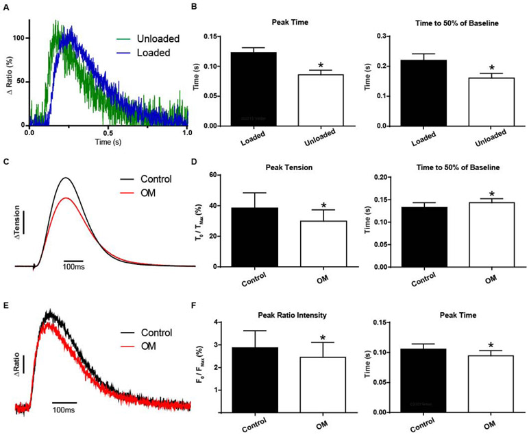
Figure 5. Load-dependent effects of myosin on sarcomere activation in live cardiac muscle.
A, Ensemble average of single myocytes (green) and papillaries (blue) reveals a load dependence to sarcomere activation (22°C and 0.2Hz). Note, FRET ratio-derived transients obtained in single myocytes were not different from Clover transient traces in the same single myocytes.
B, Summary statistics of sarcomere biosensor kinetics reveals increasing load and physiological complexity significantly delays peak activation and time to inactivation (n=8 per group). C, Ensemble average traces of tension recordings (non normalized) in papillaries before and after treatment with 250 nM Omecamtiv mecarbil shows alterations in tension development and kinetics, ΔTension bar represents a 10% change from baseline. D, Summary statistics of papillary twitch tension with 250nM Omecamtiv mecarbil reduces the change in peak tension and slows the relaxation kinetics (n=14 pairs). E, Ensemble average traces of FRET ratio recordings (non-normalized) in papillaries before and after treatment with 250 nM Omecamtiv mecarbil shows alterations in peak intensity and kinetics. ΔRatio bar represents a 5% change in FRET from baseline. F, Summary statistics of ensemble traces from e show Omecamtiv mecarbil reduces the peak FRET ratio intensity change while shortening time to peak activation (n=14 pairs). C-F, Data obtained at 22°C with 1.0Hz stimulation. Mean ± S.E.M are presented. Unpaired and paired two-tailed t-tests were used, as appropriate: *P < 0.05.

