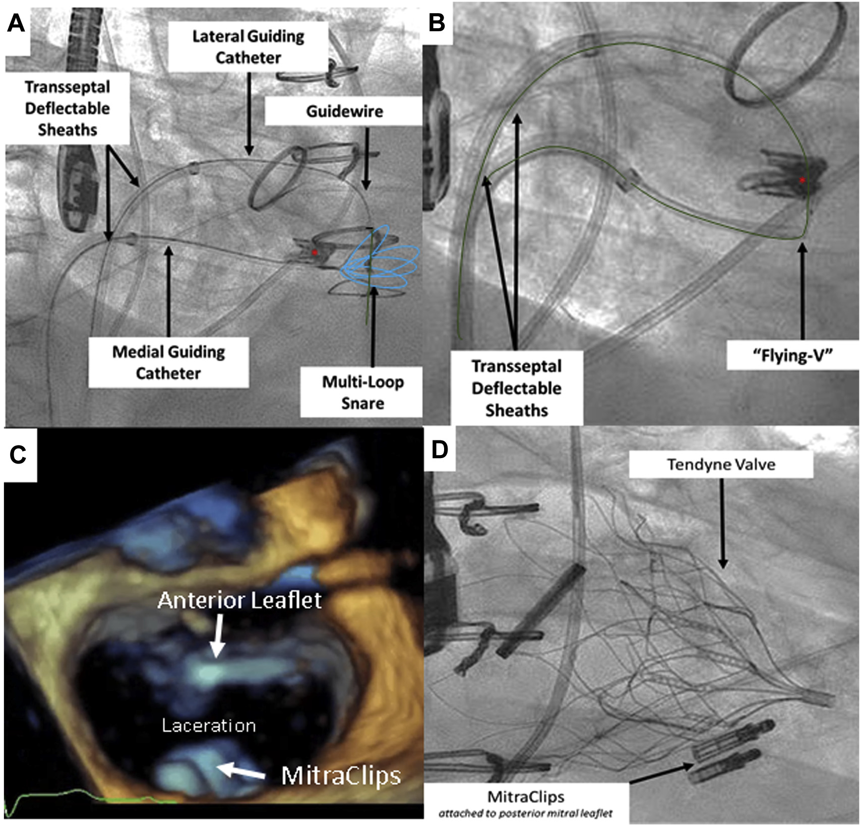
FIGURE 1. Key Steps in the Electrosurgical Laceration and Stabilization of MitraClip Procedure.

(A) After cannulation of the “double-orifice” mitral valve, an 0.014-inch guidewire (green) is advanced into a prepositioned snare (blue). Asterisk denotes MitraClip. (B) The “flying V” is created and positioned on the anterior mitral leaflet. (C) Following laceration, the singleorifice mitral valve is created. Note that the MitraClips remain attached to the posterior mitral leaflet. (D) The Tendyne valve is deployed while MitraClips remain attached to the posterior mitral leaflet.
