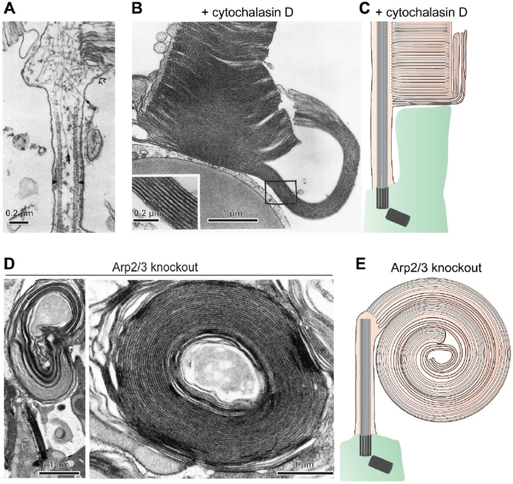
Figure 3. Branched actin polymerization initiates disc formation.

(A) An electron micrograph showing myosin-labeled actin filaments at the site of disc morphogenesis in a rat rod (adapted from [40]). (B) An electron micrograph of a cone from a Xenopus laevis retina acutely treated with the actin polymerization inhibitor cytochalasin D reveals overgrowth of newly forming discs (adapted from [43]). (C) A cartoon illustrating the effect of cytochalasin D treatment. The drug halted the initiation of new disc formation without preventing delivery of new membrane material to the outer segment. This material incorporates into the newly forming discs which were undergoing expansion at the time of treatment, causing their uncontrolled overgrowth. (D) Electron micrographs of overgrown membranes in rods of Arp2/3 knockout mice which have elaborated into large whorls. (E) A cartoon illustrating a massive membrane whorl in the Arp2/3 knockout rod. A permanent halting of the initiation of new disc formation by this knockout causes much more extended membrane outgrowth than an acute cytochalasin D treatment. Images in (C-E) are adapted from [44].
