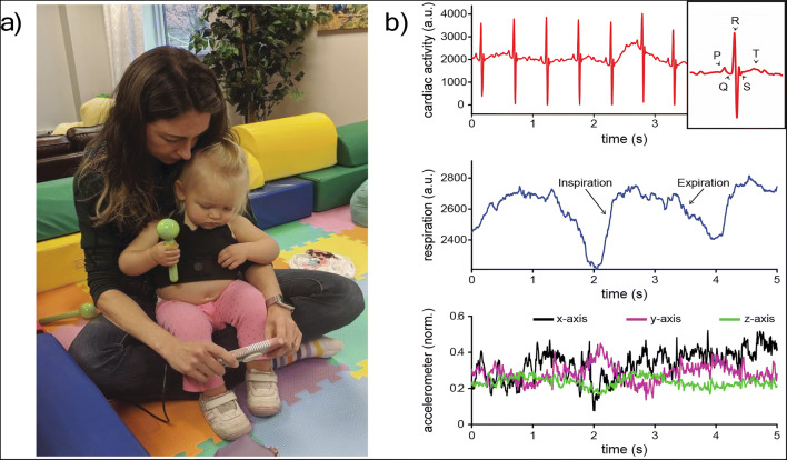
Fig. 3.
An exemplar of raw data from the autonomic vest. (a) An image of the vest applied to a 16.7-month-old child. She is sitting on her parent’s lap and watching a video on a phone, sustaining her attention to the object. (b) Five seconds of the raw data from the autonomic vest. Cardiac activity is in the top row with clear P, Q, R, S, and T waveforms identified in the inset. Respiratory activity is in the middle row with moments of inspiration and expiration identified. The third row demonstrates normalized data from the x-, y-, and z-axis of the accelerometer

