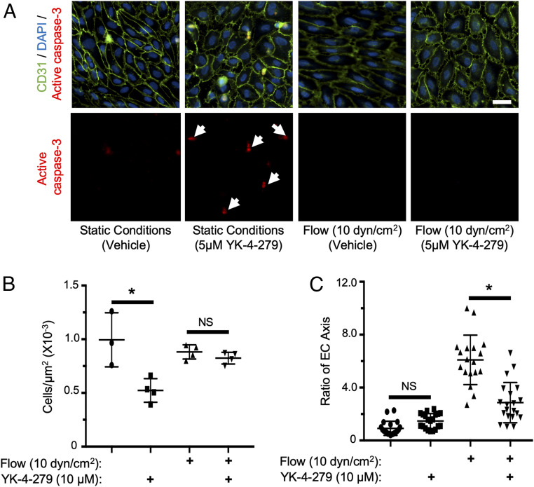
Fig. 4.
YK-4-279 induces flow-dependent HUVEC apoptosis in vitro. (A) Images of HUVECs stained for CD31 (green) and active caspase-3 (red) under the indicated flow and YK-4-279 treatment conditions; nuclei were counterstained with DAPI (blue). White arrows indicate apoptotic cells (active caspase-3+) detected after YK-4-279 treatment under static but not under flow (10 dyn/cm2) conditions. (Scale bar, 30 µm.) (B) Quantification of cells/µm2 (n = 3 to 4) using experimental conditions in A. (C) Quantification of HUVEC morphology (n = 18) as measured by the EC axis parallel to flow relative to the axis perpendicular to flow. *P < 0.05 (two-tailed Student’s t test). NS = not significant. Error bars: SD.

