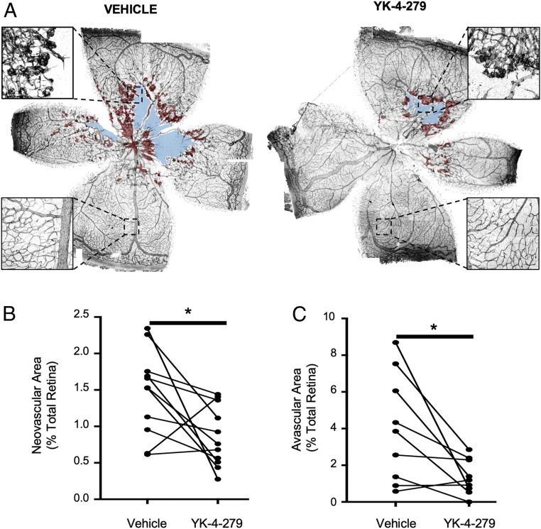
Fig. 5.
YK-4-279 reduces neovascularization and avascular areas in mouse retinas following OIR. (A) Representative images of P19.5 retinas immunostained for CD31 (black) were stitched from 50 to 60 composite images using Nikon NIS-Elements software. Retinas shown are from an individual mouse that was subjected to the OIR protocol and then intravitreally injected with YK-4-279 or a vehicle control in contralateral eyes at P17.5. Retinal neovessels and avascular areas are shaded in red and blue, respectively. (Insets) Magnifications demonstrate centrally located retinal neovessels and peripheral healthy vessels. Retinal neovascular area (B) and avascular area (C) were quantified (n = 9) and compared between YK-4-279–injected and vehicle-injected eyes. Contralateral eyes from the same mouse are indicated by the lines connecting data points. *P < 0.05 (paired two-tailed Student’s t test).

