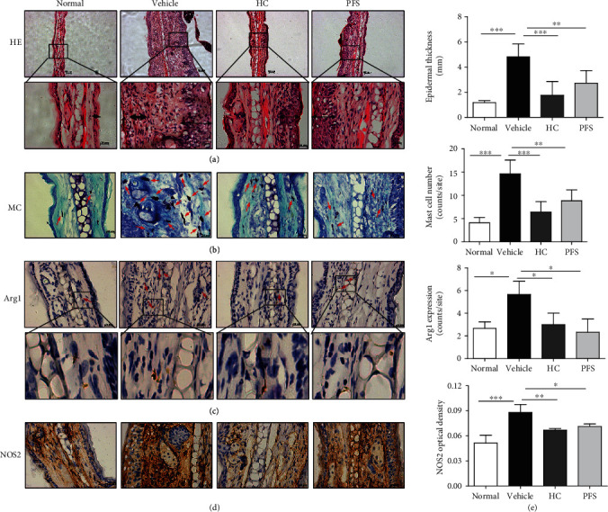
Figure 3.

PFS improved the histology of atopic dermatitis (AD) in mice (ten-time challenge). Tissues were excised, fixed in 10% formaldehyde for 1 h, embedded in OTC, and sectioned. (a) HE staining (100x; 400x); arrow indicates epidermal exfoliation; (b) toluidine blue staining (400x); arrows: mast cells; (c) Arg1 (100x; 400x); arrows: Arg1-positive macrophages; (d) NOS2 (400x); (e) epidermal thickness, number of mast cells, Arg1-positive cells, and NOS2 optical density were detected. ∗p < 0.05 and ∗∗p < 0.01 versus the vehicle group.
