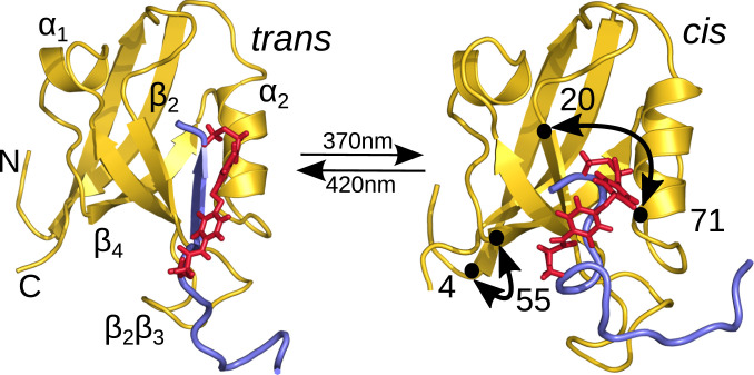
Fig. 1.
Ligand-switched PDZ2 domain. Main secondary structural elements and distances and discussed below (Results, MD Simulations) are indicated. In the trans conformation of the photoswitch (red), the ligand (blue) fits well in the binding pocket, while it starts to move out when switching to cis.

