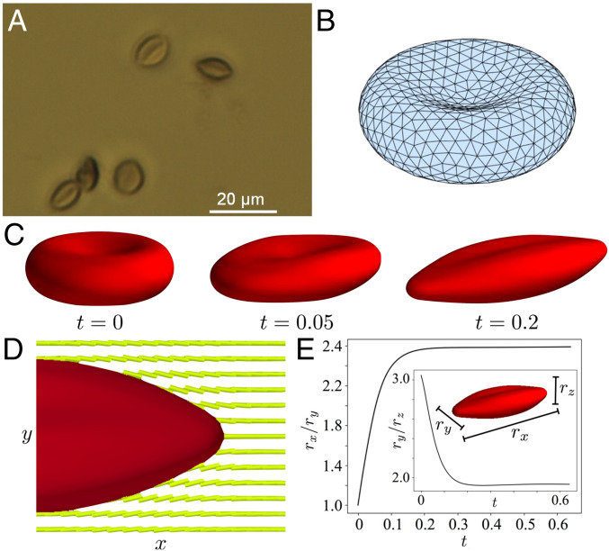
Fig. 5.
(A) Optical micrograph (63× objective) of RBCs that were cross-linked when strained by nematic LC (17.3% DSCG at 25 °C). The images were obtained after removal of the LC. (B) Triangular surface mesh of a simulated RBC in its initial biconcave configuration (prior to relaxation in LC). (C) Simulation of the RBC shape change as a function of time in nematic LC. (D) Simulated cross-section showing equilibrium RBC shape and LC configuration (Movie S2). (E) Simulated relaxation of RBC shape metrics rx/ry and ry/rz to equilibrium values when the RBC was dispersed in a LC.

