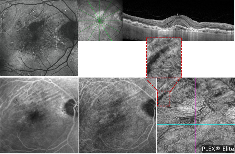
Figure 2.
Multimodal imaging from a patient with unilateral chorioretinal folds associated with macular neovascularization in right eye. (Row above): (Left) blue fundus autofluorescence (BAF) image shows linear folds at the macula, easier to detect in the superior region. Structural optical coherence tomography (OCT) image (right) confirms the presence of CFRs. (Row below): The late fluorescein angiography (FA - left) and intermediate indocyanine green angiography (ICGA - middle) images show typical alternating hypofluorescent and hyperfluorescent bands. The optical coherence tomography angiography (OCTA - right) image shows a transversal line of signal reduction corresponding to vascular rarefaction at the choriocapillaris layer.

