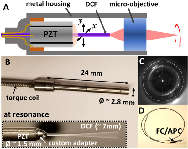
Fig. 1.
A, a schematic of a resonant fiber-optic scanning endomicroscope. A precision-machined PZT base and a custom adapter were used for centering each component and ensuring effective force transfer from the PZT actuator to the fiber cantilever. B, photograph of a fully assembled endomicroscope. The fiber scanner and a superachromatic micro-objective are encased inside a hypodermic metal tube of a 2.8 mm diameter and a 24 mm overall rigid length. A ~7 mm long DCF was cantilevered at the distal end of a PZT actuator and resonantly scanned (inset). C, front view of an endomicroscope showing the DCF was concentrically aligned with the longitudinal axis of the endomicroscope. D, photograph of an assembled endomicroscope with a protective torque coil over the fiber and PZT drive wires and an FC/APC connector at its proximal end.

