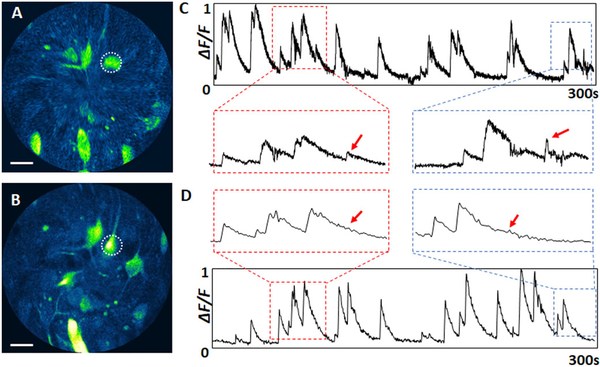
Fig.4.
A and B, representative MPF images of neuronal activities in the primary motor cortex of a freely-behaving rodent captured with the endomicroscope at imaging speeds of A, 26.4 fps and B, 3.3 fps. C and D, recorded calcium dynamics of a neuron indicated with a dotted circle in A and B over 5 min: Cat 26.4 fps, Dat 3.3 fps. Scale bars: 20 μm.

