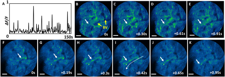
Fig. 5.
Dendritic activities of a freely-behaving mouse captured with the endomicroscope. A, calcium dynamics of a dendrite indicated with a white arrow captured at 26.4 fps. B–K, time-series images of a dendrite burst captured at B–E, 3.3 fps and F–K, 26.4 fps. The blood vessels are indicated with yellow arrows in B, and similar blood vessels can be seen in all the other images. Scale bars: 20 μm.

