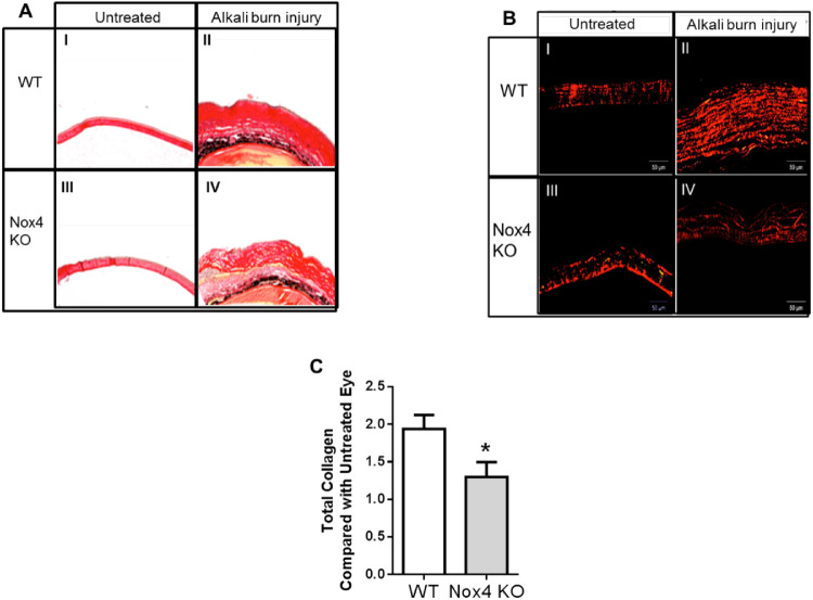
Figure 5.
Collagen fibers in mouse cornea detected by picrosirius red polarization method. Collagen staining of WT and Nox4 KO corneas shows an increase of collagen fibers after NaOH treatment (II and IV, respectively) compared with untreated control (I and III, respectively) using (A) nonpolarized light (4×) and (B) polarized light (10×). (C) Quantitation shows a significant reduction of collagen ratio after NaOH treatment in Nox4 KO-derived compared with WT-derived cornea. Values (mean ± SEM; n = 4–5) are represented as a fold change from WT control; *P < 0.05 from WT following a Student's paired t-test. Scale bar: 50 µm.

