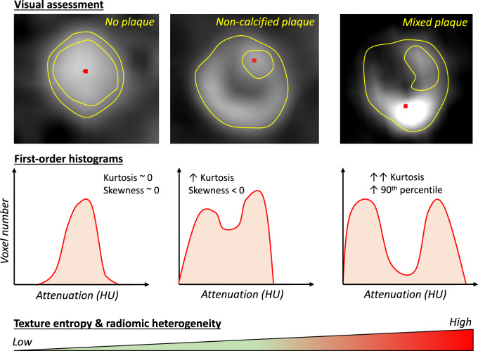
Figure 3.
Radiomic phenotyping of coronary lesions. Differences in coronary plaque composition will manifest as different radiomic texture patterns on computed tomography analysis, which can then be quantified using first- and higher-order radiomic features. Changes in these metrics can be used in an automated way to not only detect plaques but also produce a deep characterization of the histology and biology of a given lesion.

